INFRAROSSO (IR)
L’infrarosso (IR) è una lunghezza d’onda molto lunga che va dal limite del visibile (720nm) a 1350nm; per inciso: i limiti di cui sopra rappresentano gli estremi che più interessano in fotografia, lunghezze maggiori fino a 1mm non ci interessano. La luce infrarossa è invisibile all’occhio ma non a pellicole (una volta ne esistevano diverse per questo tipo di riprese) e a sensori CCD/CMOS etc.
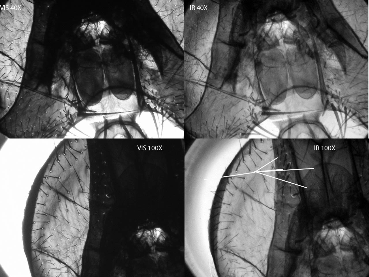
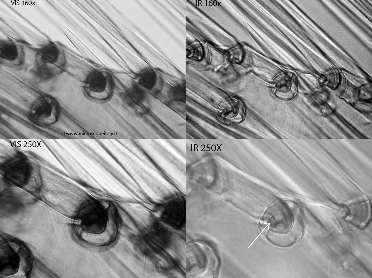
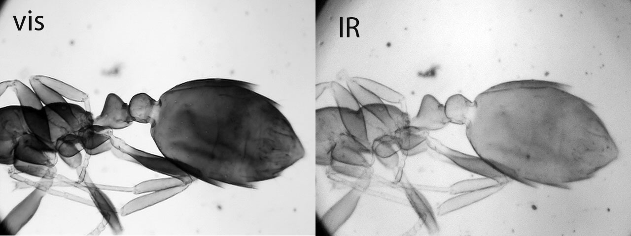
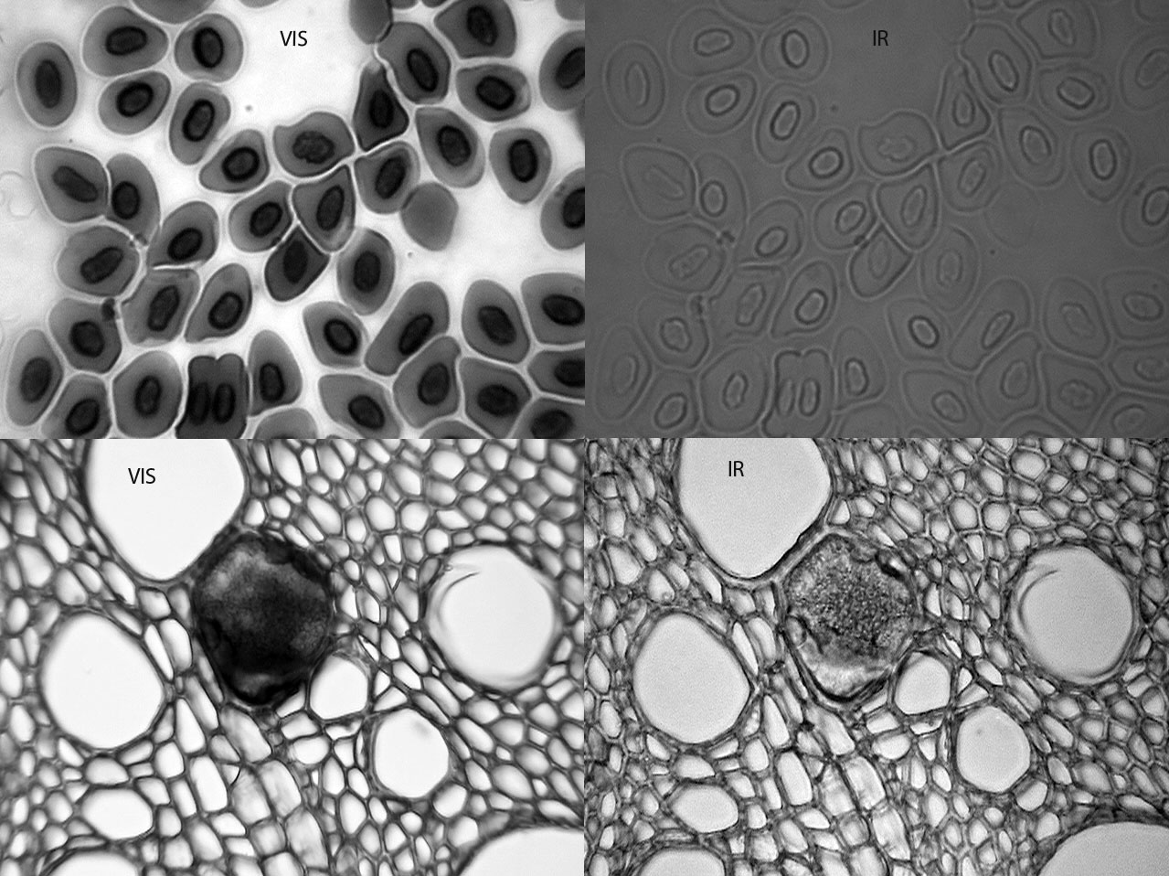
Ovviamente le luce IR può essere adoperata in microscopia con risultati degni di nota. Giusto per conoscenza, il primo ad utilizzarla fu August Kohler nel 1891. Il vantaggio di questa radiazione è che materiali normalmente opachi se osservati in luce normale trasmessa, risultano più trasparenti se osservati con luce IR; così ad esempio, la chitina degli insetti, la clorofilla, campioni densamente colorati, fibre tessili etc., mostreranno nuovi particolari. Senza entrare nello specifico dei grossi microscopi progettati per l’IR, tutto ciò che ci serve è:
Filtro IR: meglio quelli a 720nm, che con normali microscopi vanno più che bene essendo leggermente più trasparenti di quelli da 950nm, almenochè non si disponga di illuminazioni molto intense. Si trovano in quantità sul web.
Camera/telecamera/webcam: senza filtro “IR cut” o con possibilità di inserirlo e disinserirlo al bisogno
La telecamera che possiedo e che mi da ottimi risultati, è una Sony DCRTRV80E, la quale monta un selettore per inserire e disinserire l’”IRcut” caratteristica questa di molti prodotti Sony ; è ovvio che questo casca a pennello per gli scopi di cui sopra.
Chi non ha una camera specifica, senza svenarsi può utilizzare una webcam (meglio megapixel) a patto che si rinunci un po’ alla qualità e si prenda in considerazione l’idea di smontarla per rimuovere l’ IR cutche di solito si presenta come una sottile lamina di vetro rosato, posta esattamente davanti al sensore!
Anche con un’attrezzatura non proprio specialistica, quindi, è possibile trarre soddisfazione nell’osservare campioni o particolari di essi, che prima erano invisibili!
NOTE: Per quanto riguarda la fotomicrografia, non vi sono particolari accorgimenti se non quello che il campione necessita di una nuova messa a fuoco, infatti ciò che è perfettamente a fuoco nel visibile non lo sarà nell’IR e bisognerà correggere!
Devo anche aver letto da qualche parte che l’IR è dannoso per gli occhi, si eviti dunque, di osservare nell’oculare (anche perché non serve a nulla) e ci si basi solo sulle immagini fornite dal sistema elettronico!
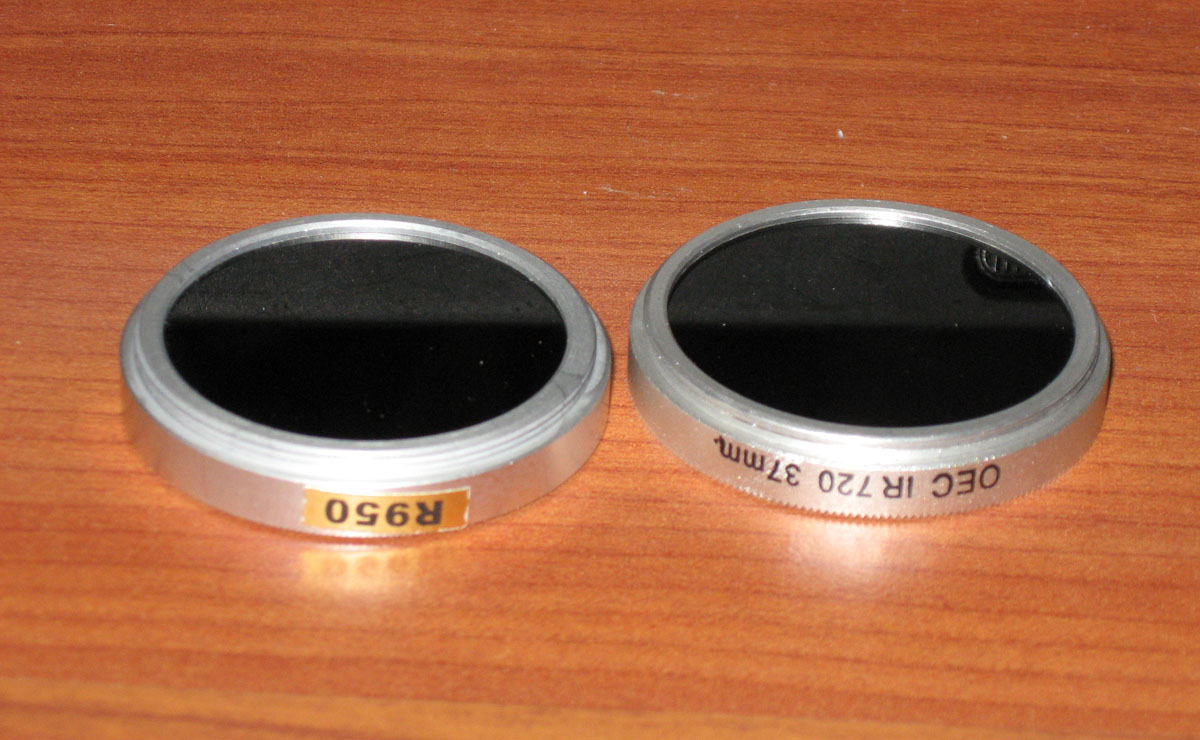 Filtri - filters
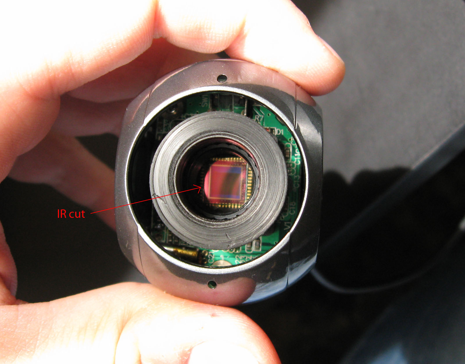 IR cut
 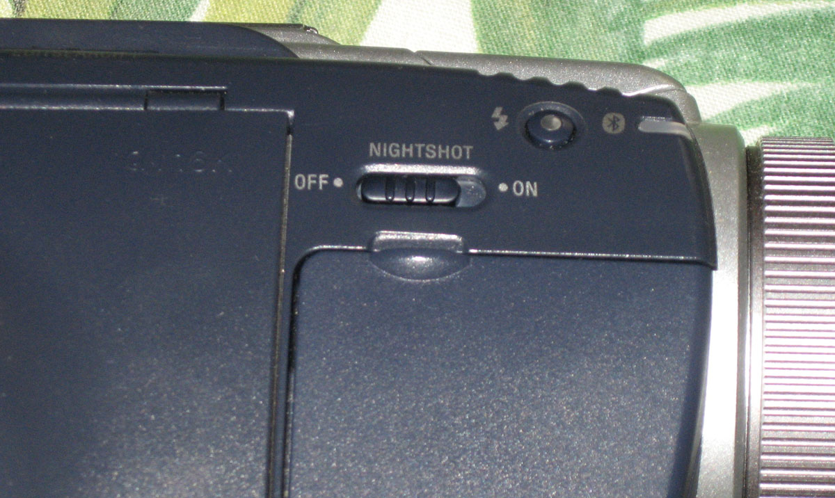 selettore IR su Sony/ Sonycam IR selector
The infrared (IR) is a very long wavelength that goes from the edge of the visible (720nm) to 1350nm, incidentally; the limits mentioned above represent the extremes of interest in photography, longer lengths, up to 1 mm does not interest microscopysts. Infrared light is invisible to the eye but not to film and CCD / CMOS sensor.
Obviously, the IR light can be used in microscopy with remarkable results. Just to know, August Kohler was the first to use IR in 1891. The advantage of this radiation is that material normally opaque when viewed in normal transmitted light , are more transparent when viewed with IR light, so for example, chitin of insects, chlorophyll, densely colored samples, textiles etc.., show new details . Without entering into the big microscopes designed for IR, all we need is:
IR filter: better 720nm, which with ordinary microscopes are just slightly more transparent than those from 950nm.There are a lot on the web.
Camera / camcorder / webcam: whitout “IR cut”
The camera I own and that gives me good results, is a Sony DCRTRV80E, which has a switch for activating and deactivating the ‘IRcut “..thi is a characteristic of many Sony products (see above).
If you have not a camera, you can use a webcam (better megapixels) but you have to take into consideration the idea to disassemble it to remove the’ IR cut that usually presents as a thin sheet of glass rose (see above), placed exactly on the sensor!
Then, even without specialist equipment , you can get satisfaction in observing samples or details of them, invisible before!
NOTE: For photomicrography, there are no special precautions except that the sample requires a new focus, because what is perfectly in focus in normal lighting will not be visible in the IR and you have to correct fine focusing!
I also have read somewhere that the IR is harmful to the eyes, avoid therefore, to observe in the eyepiece and use only on the images provided by the electronic camera.
Alcuni esempi/some exemple